 What is the knee joint?
- What causes knee pain?
- What is the shoulder joint?
- What causes shoulder pain?
- Can physical therapy help my knee and shoulder pain?
Joint pain is common in both the knees and the shoulder. One study found that the spread of joint pain is not random. Researchers discovered that persistently painful knees also seem to be correlated to bilateral shoulder pain. The study determined, “biomechanical factors influence the spread of pain.”
Both the knees and the shoulders can suffer from the same kinds of injuries or conditions, including:
- Arthritis
- Broken bones
- Wear and tear injuries
Your bones, cartilage, joints, ligaments, muscles, and tendons all work together to keep you upright and moving forward. However, when arthritis, a form of joint inflammation, crops up in the body in one place, the chances are you’ll feel it elsewhere, too. That’s why knee pain can throw the rest of the body off, causing pain in other joints, including your shoulders.
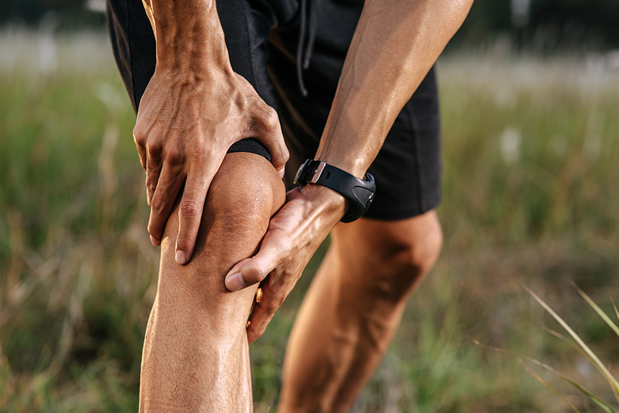
What is the Knee Joint?
 The knee is the largest and most complicated weight bearing joint structure in the body. It plays a critical role in human mobility. This joint serves to:
- Act as a stabilizer and shock absorber
- Help raise or lower the body
- Let the leg twist and move laterally
- Make walking more efficient
- Propel the forward motion of the body
- Support the upright position of the body
Three bones come together at the knee joint; the femur (upper leg bone), tibia (lower leg bone), and the patella (kneecap). All are held together by muscles, tendons, and ligaments.
The knee is a synovial joint, which means it is filled with fluid that lubricates the smooth sliding of the bones. There are 14 bursa, or small fluid-filled sacs within the joint. They help cushion and reduce friction while preventing inflammation. Two types of cartilage in the knee serve to keep the joint cushioned as you apply stressors in the form of running, climbing stairs, squatting, and even walking. It’s no wonder the knee is so commonly injured during sports or other physical activities.
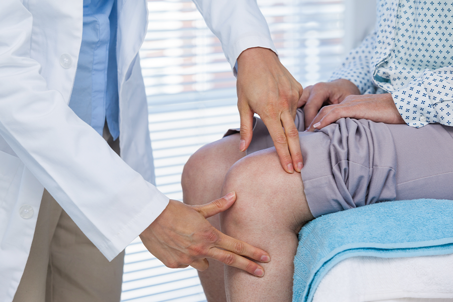
What Causes Knee Pain?
 Pain in the knee joint is one of the most common complaints of people of all ages and from all walks of life. Knee pain can stem from injury, arthritis, or mechanical problems in the joint.
Knee injuries could stem from damage to one or more of the myriad components that make up the knee, including:
- Anterior cruciate ligament (ACL)
- Bursitis
- Meniscus
- Tendinitis
Strains and sprains within the knee ligaments are common. These injuries happen during physical activity, typically related to a sudden twisting, a rapid change in direction, or a jump where the person landed wrong. The knee joint or the bones around the knee can also be fractured. For example, a patella fracture could happen from a car or motorcycle accident, or from a kick in the knee during a sporting event. Injury can also occur from overusing the joint.
Arthritis can form from an old injury or the joints can grow inflamed from overuse. There are more than 100 different types of arthritis that can affect your joints, including two of the most common, osteoarthritis and rheumatoid arthritis.
Mechanical problems in the knee can cause a lot of pain and lead to other issues like arthritis and a lack of mobility. The cause of these problems can vary. For example, an injury can cause a piece of cartilage or bone to break off and float until it gets caught in the joint. Your knee could also be dislocated and slip out of place.
The knee joint has a critical job to do. However, there is another joint in the body that gets just as much wear and tear. Let’s look at the shoulder joint and what it does to keep you moving.
What is the Shoulder Joint?
 While most people think of the shoulder as a single joint, there are actually four joints that contribute to the shoulder:
- The glenohumeral joint is what most of us think of the ball and socket shoulder joint
- The acromioclavicular joint is where the collarbone (clavicle) glides along the scapula
- The sternoclavicular joint is where the clavicle meets the top of the chest
These joints serve to bring together the humerus (upper arm bone), the scapula (shoulder blade), and the clavicle (collar bone). These bones have a cartilage cushion and four tendons called the rotator cuff. There are also muscles and ligaments that tie everything together. The shoulder also has bursa to cushion these moving parts. Like the knee, the synovial membrane produces fluid to lubricate the joint.
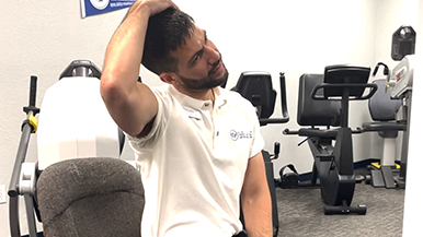
What Causes Shoulder Pain?
 Several conditions can lead to shoulder pain. Some of the most common include:
- Arthritis
- Bone spurs
- Broken bone
- Dislocated shoulder
- Heart attacks
- Inflamed tendons
- Pinched nerve
- Repetitive use injuries
- Spinal cord injuries
- Swollen bursa sacs
- Torn cartilage
- Torn rotator cuff
Depending on the condition, your doctor may prescribe anything from medications to massage, physical therapy or surgery. But when you have both bad knees and bad shoulders, what are your options?
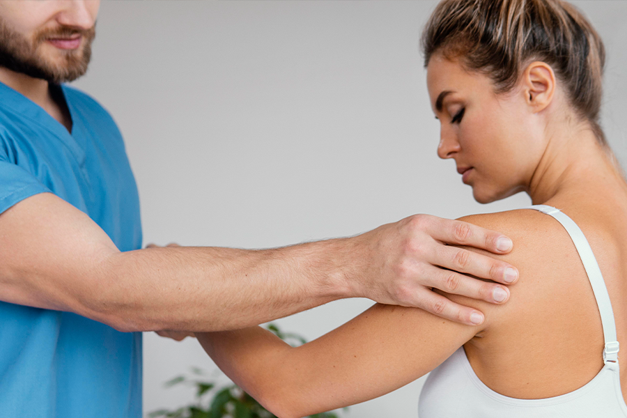
Can Physical Therapy Help My Knee and Shoulder Pain?
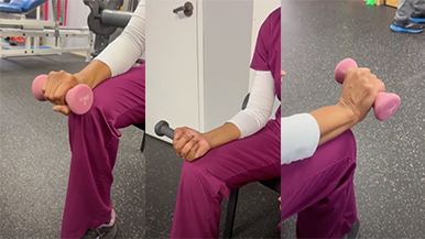
A physical therapist can work with you to develop a rehabilitation plan to improve the functionality and lessen the pain of your knees and shoulders. Most rehab plans include:
- Braces to restrict joint movement as it heals
- Exercises to strengthen the muscles around the joint
- Passive range of motion exercises to restore joint flexibility
Ability Rehabilitation can help you lessen your pain, heal, and manage symptoms and mobility – all without a physician referral. If you’re experiencing pain, schedule a consultation with one of our joint pain experts to see how you can avoid surgery and experience relief today.